SRL総合検査案内 TEST DIRECTORY

病理学的検査 - 細胞診検査

pathological test - cytodiagnosis

2024/08/09→大原 2018/12/14 2019/04/22 2019/04/22 2019/04/22 2019/04/22 2019/04/22 - 2022/02/09 2022/06/15 2019/04/22 2024/08/09 - 未確認 2024/04/18

細胞診検査注記

依頼書、スライドグラスおよびオブジェクトケースへの記入方法

依頼書

細胞診検査は各種の臨床情報を参考にして行う形態学的検査です。 細胞診検査のご依頼にあたっては専用の細胞診検査依頼書に下記の内容を記載してください。 細胞診の判定内容を左右する因子ですので記載もれがないようお願いいたします。

  • a) 氏名、性別、年齢、生年月日、科名等
  • b) 検査材料、採取方法
  • c) 臨床診断、臨床経過、検査、治療諸事項
  • d) 病変の大きさ、性状
  • e) 婦人科材料や乳腺材料の場合月経等諸事項
  • f) 前回の登録標本番号
  • g) ご提出のスライドグラス枚数

スライドグラス

  • a) スリガラス部に氏名をご記入ください。
  • b) 乾燥固定の場合は「乾燥」または「ドライ」とご記入ください。

オブジェクトケース

  • a) 氏名、性別、年齢
  • b) 標本枚数 (湿固定枚数と乾燥固定枚数)

依頼書等への記入方法

依頼書

細胞診検査は各種の臨床情報を参考にして行う形態学的検査です。 細胞診検査のご依頼にあたっては、専用の細胞診検査依頼書に下記の内容を記載してください。 特に「臨床所見および経過」欄は細胞診の判定内容を左右する因子ですので必ずご記入ください。
依頼書は黒ボールペンで強くご記入ください。

  • a) 患者名、性別、生年月日 (必ずご記入ください。) 採取日時、年齢、科名等
  • b) 検査材料、検体本数、標本枚数
  • c) 臨床診断、臨床経過、治療諸事項
  • d) 病変の大きさ、性状
  • e) 婦人科材料や乳腺材料の場合、月経等諸事項
  • f) 前回結果
  • g) 前回の登録標本番号

スライドグラス

  • a) スリガラス部に患者名をご記入ください。
  • b) 乾燥固定の場合は「乾燥」または「ドライ」とご記入ください。
  • c) 材料名をご記入ください。

オブジェクトケース

固定された標本をオブジェクトケースに入れてご提出ください。 また、細胞診検査依頼書に添付されている検体ラベルに採取日、患者名、性別、年齢を明記してオブジェクトケースにお貼りください。

検体容器

検体を入れてご提出ください。 また、細胞診検査依頼書に添付されている検体ラベルに採取日、患者名、性別、年齢を明記して検体容器にお貼りください。 (貼り方:右図参照)

ラベル

(注意) 細胞診検査は、必ずしも確定診断ではありません。 診断はご提出いただいた標本において下されたもので、病変全体を反映するものではないことを予めご了承ください。 組織診検査等による総合的な診断をお勧めします。(検診検体は偽陰性となる可能性があります)

依頼不可検体

検体の取り扱い上、以下の検体についてはご依頼できませんので、ご注意ください。

  • ホルマリン固定された検体
  • 凍結保存された検体
  • 修復不可能な破損標本
  • 右記に示すような検査材料 組織、爪、毛髪、便、 血液、胆石、結石、骨髄、胃液、シードスワブ

検査材料の提出方法 [塗抹、固定、検査材料、検体量]

各種材料の塗抹

塗抹の仕方は検査材料の種類、性状などに応じて異なります。下記の要領で塗抹してください。

標本の固定

固定は細胞の変性・融解などの変化を停止させる作業ですので、塗抹後直ちに固定する必要があります。 湿固定はパパニコロウ染色、PAS染色などのための固定法で、乾燥固定はギムザ染色などのための固定法です。 固定の良否は採取部位・方法とともに細胞診判定内容を左右する重要な因子の一つです。迅速な固定をお願いいたします。

  1. 湿固定【婦人科細胞診・一般細胞診細胞診 (その他)】

    塗抹したスライドグラスを95%エタノールに30分以上浸漬して固定してください。湿固定にはサイトタック等を用いたコーティング固定法もあります。浸漬後、ラビッドスプレー等を用いてコーティングしてください。 コーティング固定剤を使用する場合は、ムラが生じないよう固定液を塗抹全面に充分に噴霧または滴下してください。

  2. 乾燥固定【一般細胞診細胞診 (その他)】

    塗抹後直ちに、塗抹面を扇風機や冷風ドライヤーなどで急速に乾燥させます。 自然乾燥は乾燥むらを生じるので不充分です。

■検査材料、検体量

【一般細胞診細胞診 (その他) [項目コード:00541 0] [項目コード:0541 8] [項目コード:8029] [項目コード:871237]】
  1. 喀痰
    「小豆」大の喀痰を2枚のスライドグラスの間にはさみ、前後左右に押しつぶすように満遍なくのばしてください (すり合わせ法)。 癌細胞は血痰部や不透明白濁部、ゼリー状粘液部に多く含まれるので、その部分を塗抹してください。
    検体量:湿固定塗抹標本 2枚
    また、生材料は、フィブリンの析出を防ぐために抗凝固剤入り容器で数本採取してよく混和し、10mLをポリスピッツに移してご提出ください。
    検体量:生材料10mL
  2. 擦過材料 (気管支、消化管、胸膜・腹膜等)
    ブラシ、綿棒等により病巣などを擦過しスライドグラスに塗抹してください。 擦過物は非常に乾燥しやすいので塗抹固定を迅速に行うことが必要です。
    検体量:湿固定塗抹標本 1枚、乾燥固定塗抹標本 1枚
  3. 液状材料 (尿、胸水、腹水、心嚢液、陰嚢水腫液、各種洗浄液等)
    1500rpm5分間遠心後、沈渣をすり合わせ法または引きガラス法によって塗抹してください。 血性検体の場合は有核成分が多いバフィーコート部分を塗抹してください。
    検体量:尿・胸水・腹水・心嚢液・陰嚢水腫液、各種洗浄液等  湿固定塗抹標本 12枚、乾燥固定塗抹標本 1枚
  4. 針穿刺吸引材料 (乳腺、甲状腺、肺、リンパ節、前立腺、睾丸、軟部腫瘍、肝等)
    穿刺針内に吸引された材料をスライドグラス中央に静かに吹きだし塗抹します。 検体が微量なときはもう1枚のスライドグラスを合わせてからはがし2枚作製してください。 多めに得られたときや多量の場合は引きガラス法で塗抹してください。
    検体量:湿固定塗抹標本 12枚、乾燥固定塗抹標本 1枚
  5. 捺印標本 (各種固定腫瘍、リンパ節等)
    メスやカミソリで切った新しい組織割面をスライドグラスに軽く捺印してください。
    検体量:湿固定塗抹標本 1枚、乾燥固定塗抹標本 1枚
  6. 圧挫標本 (中枢神経腫瘍、甲状腺腫瘍等)
    組織の小塊 (直径1-2mm) を2枚のスライドグラスの間にはさみ、軽く押しつぶして組織が伸展したらスライドグラスをはなし、 厚い方を湿固定、薄い方を乾燥固定してください。
    検体量:湿固定塗抹標本 1枚、乾燥固定塗抹標本 1枚
  7. 蓄痰材料
    細胞診 [喀痰集細胞] 専用容器に蓄痰し、容器ごと提出してください。
    細胞診 [喀痰集細胞] 専用容器の取り扱い方法

    ● 1日1回早朝痰を専用容器に喀出し、痰を入れたらフタをして15~20回強く振ってください。 この操作を3~5日連続して行います。 痰の出にくいときは、茶碗等に熱湯を満たしその蒸気を吸いながら5~6回大きく深呼吸すると痰は出やすくなります。

  1. 擦過材料 (気管支、消化管、胸膜・腹膜等)
    ブラシ、綿棒等により病巣などを擦過しスライドグラスに塗抹してください。 擦過物は非常に乾燥しやすいので塗抹固定を迅速に行うことが必要です。
    検体量:湿固定塗抹標本 1枚、乾燥固定塗抹標本 1枚
  2. 針穿刺吸引材料 (乳腺、甲状腺、肺、リンパ節、前立腺、睾丸、軟部腫瘍、肝等)
    穿刺針内に吸引された材料をスライドグラス中央に静かに吹きだし塗抹します。 検体が微量なときはもう1枚のスライドグラスを合わせてからはがし2枚作製してください。 多めに得られたときや多量の場合は引きガラス法で塗抹してください。
    検体量:湿固定塗抹標本 1枚、乾燥固定塗抹標本 1枚
  3. 捺印標本 (各種固定腫瘍、リンパ節等)
    メスやカミソリで切った新しい組織割面をスライドグラスに軽く捺印してください。
    検体量:湿固定塗抹標本 1枚、乾燥固定塗抹標本 1枚
  4. 圧挫標本 (中枢神経腫瘍、甲状腺腫瘍等)
    組織の小塊 (直径1-2mm) を2枚のスライドグラスの間にはさみ、軽く押しつぶして組織が伸展したらスライドグラスをはなし、 厚い方を湿固定、薄い方を乾燥固定してください。
    検体量:湿固定塗抹標本 1枚、乾燥固定塗抹標本 1枚
<注意>

検体採取から塗抹固定までの許容時間 (室温)

  • 喀痰・胸水・腹水・心嚢液・・・12時間以内
  • 尿・穿刺液・洗浄液・髄液・・・1時間以内
  • 胆汁・膵液・十二指腸液・・・氷冷中の容器に採取し、1時間以内
  • 擦過材料・捺印材料・圧挫材料・・・5秒以内
【細胞診 (婦人科) [項目コード: 00695 00695 8]、 細胞診 (婦人科ベセスダシステム) [項目コード: 06242 66242 5]】 【婦人科細胞診 [項目コード:8030]】 【細胞診婦人科〔直接塗抹法〕 [項目コード:871137]】
  • 婦人科材料

    乾燥しやすいので塗抹後直ちに湿固定してください。
    検体量:湿固定塗抹標本 1枚

●検体の取り扱い上、以下の検体についてはご依頼できませんので、ご注意ください。

  • ホルマリン固定された検体
  • 凍結保存された検体
  • 修復不可能な破損標本
  • 下記に示すような検査材料
    組織、爪、毛髪、便、血液、胆石、結石、骨髄

検査結果の報告

1. ベセスダシステム

標本の適正・不適正の評価について

適正検体:保存状態がよく、鮮明に見える扁平上皮細胞が直接塗抹法では8000~12000個、LBCの場合では5000個以上とされています。
不適正検体:扁平上皮細胞の75%以上が不明瞭である場合、 炎症細胞、出血により覆われていた場合、過度な乾燥によるアーチファクトが見られた場合などがあてはまります。
*適正検体とされた場合に、下記の細胞判定が実施されます。

ベセスダ表記 表記の説明 推定病変
陰性 (NILM) 陰性 微生物
その他の非腫瘍性所見
扁平上皮系異常
ASC-US 意義不明な異型扁平上皮細胞 軽度扁平上皮内病変疑い
ASC-H HSILを除外できない異型扁平上皮細胞 高度扁平上皮内病変疑い
LSIL 軽度扁平上皮内病変 HPV感染
軽度異形成
HSIL 高度扁平上皮内病変 中等度異形成
高度異形成
上皮内癌
SCC 扁平上皮癌 扁平上皮癌
腺系異常およびその他の悪性腫瘍
AGC 異型腺細胞 腺異型または腺癌疑い
AIS 上皮内腺癌 上皮内腺癌
Adenocarcinoma 腺癌 腺癌
Other malig. その他の悪性腫瘍 その他の悪性腫瘍

2. Class判定

Class Ⅰ
: Abscence of atypical or abnormal cells.
Class Ⅱ
: Atypical cytology but no evidence of malignancy.
Class Ⅲ
: Cytology suggestive of, but not conclusive for malignancy.
Class Ⅲa
: Probably benign atypia.
Class Ⅲb
: Malignancy suspected.
Class Ⅳ
: Cytology strongly suggestive of malignancy.
Class Ⅴ
: Cytology conclusive for malignancy.

*Class判定について、Ⅰ、Ⅱを陰性、Ⅲ、Ⅲa、Ⅲbを疑陽性、Ⅳ、Ⅴを陽性と読み換えることが可能です。

判 定 パパニコロウ分類 細胞診所見
陰性
(−)
Class Ⅰ 異型細胞をみない
Abscence of atypical or abnormal cells.
Class Ⅱ 異型細胞はあるが、悪性細胞をみない
Atypical cytology but no evidence of malignancy.
疑陽性
(±)
Class Ⅲ 悪性を疑わせる細胞をみるが確診できない
Cytology suggestive of, but not conclusive for malignancy.
Ⅲa Probably benign atypia.
Ⅲb Malignancy suspected.
陽性
(+)
Class Ⅳ 悪性、極めて濃厚な異型細胞
Cytology strongly suggestive of malignancy.
Class Ⅴ 悪性と診断可能な異型細胞
Cytology conclusive for malignancy.

* 婦人科細胞診で検査材料が子宮内膜の場合は、上表の判定の内容にて結果をお返しいたします。

標本の保管時間

陰性 (NILM)、ClassⅠ・Ⅱの診断標本は5年間保管し、ASC-US以上、ClassⅢa以上のものは10年保管いたします。

3. ABC判定

判定区分 細胞所見 指導区分
A 喀痰中に組織球を認めない 材料不適、再検査
B 正常上皮細胞のみ
基底細胞増生
軽度異型扁平上皮細胞
繊毛円柱上皮細胞
現在異常を認めない
次回定期検査
C 中等度異型扁平上皮細胞
核の増大や濃染を伴う円柱上皮細胞
再塗抹または6ヵ月内の再検査
D 高度 (境界) 異型扁平上皮細胞または悪性腫瘍が疑われる細胞を認める 直ちに精密検査
E 悪性腫瘍細胞を認める
肺癌検診における喀痰細胞診の判定基準と指導区分 (2016改定) 臨床・病理肺癌取扱い規約第8版

参考文献

  • Masayoshi Takahashi: Color Atlas of Cancer Cytology. IGAKU-SHOIN: 1981.
  • 田中 昇,他:細胞診教本.宇宙堂八木書店:1988.
  • 田嶋 基男,他:細胞診全書II細胞診の基本技術および細胞診に必要な病理学各論.宇宙堂八木書店:1980.
  • 栗原 操寿,他:日本臨床細胞学会教育委員会,編:臨床細胞診断学教本.医学図書出版:1985.
  • 高橋 正宜,他:病理と臨床3 (臨時増刊号) 針生検細胞診.文光堂:1985.
  • 日本病理学会,編:病理技術マニュアル6 細胞診とその技術.医歯薬出版:1982.
  • 上井 良夫:病理と臨床7 (臨時増刊号) 腫瘍細胞診 最近の進歩:1989.
  • 矢谷 降一,他:細胞診を学ぶ人のために.医学書院:1990.
  • D.Solomon, R.Nayar編 平井康夫監訳:ベセスダシステム2001アトラス
  • Ritu Nayar,David C.Wilbur編 平井康夫監訳:ベセスダシステム2014アトラス 子宮頸部細胞診報告様式:2016.
  • 日本産婦人科医会:ベセスダシステム2001準拠子宮頸部細胞診報告様式の理解のために:2008.12
  • 細胞診を学ぶ人のために. 医学書院
  • 細胞診の基本技術および細胞診に必要な病理学各論. 宇宙堂八木書店
  • 細胞診のすすめ方. 近代出版
  • スタンダード細胞診テキスト. 医歯薬出版
  • 細胞診の基本. 武藤化学株式会社出版
  • 細胞診カラー図鑑. HBJ出版局
  • 尿中細胞アトラス. 医歯薬出版
  • 体腔液細胞診アトラス. 篠原出版新社
  • Thinlayer標本アトラス 婦人科細胞診. MBL 株式会社医学生物研究所
  • ベセスダ・システム2001アトラス. 丸善出版
  • ベセスダ・システム2001準拠子宮頸部細胞診報告様式. 日本産婦人科医会提唱
  • ベセスダ・システムの基礎と実践-その理解のために-. 武藤化学株式会社出版
  • 臨床検査 51巻8号. 医学書院
  • 石和 久,他:Medical Technology 35(10):1077~1080, 2007.
  • 岩坂 剛,他:産科と婦人科 73(2):197~203, 2006.
  • 産婦人科診療ガイドライン-婦人科外来編2011. 日本産科婦人科
  • 子宮頸がん検診とヒトパピローマウイルスQ&A. 日本細胞診断学推進協会
  • 記述式内膜細胞診報告様式に基づく 子宮内膜細胞診アトラス. 医学書院
  • 実践細胞診テキスト 初心者からエキスパートまで. 大阪大学出版会
  • 新版 乳腺細胞診カラーアトラス-乳癌取扱い規約「細胞診報告様式」に準拠. 医療科学社
  • 遺伝子検査関連 検体品質管理マニュアル. 日本臨床検査標準協議会(JCCLS)
  • 遺伝子検査関連 検体品質管理マニュアル(パート2). 日本臨床検査標準協議会(JCCLS)
  • 液状化検体細胞診断マニュアル. 篠原出版新社
  • 細胞診ガイドライン1 婦人科・泌尿器. 金原出版
  • 細胞診ガイドライン2 乳腺・皮膚・軟部骨. 金原出版
  • 細胞診ガイドライン3 甲状腺・内分泌・神経系. 金原出版
  • 細胞診ガイドライン4 呼吸器・胸腺・体腔液・リンパ節. 金原出版
  • 細胞診ガイドライン5 消化器. 金原出版
  • 子宮頸部細胞診運用の実際 第2版:ベセスダシステム2014準拠. 医学書院
  • ベセスダシステム2014アトラス 子宮頸部細胞診報告様式. 丸善出版
  • 矢谷 降一,他:細胞診を学ぶ人のために. (第4版) 医学書院:2005.
  • D.Solomon, R.Nayar編 平井康夫監訳:ベセスダシステム2001アトラス
  • 日本産婦人科医会:ベセスダシステム2001準拠子宮頸部細胞診報告様式の理解のために:2008.12
  • 西 国広,編:〜基礎から学ぶ〜細胞診のすすめ方(第4版)近代出版:2018.

Notes for cytodiagnosis test

How to fill out the request form, glass slides and object case

Request form

A cytodiagnosis test is a morphological test performed with reference to various clinical information. When requesting a cytodiagnosis test, please fill out the following information on the dedicated cytodiagnosis test request form.
Please be sure to fill all the requested information, as it is a factor that influences the cytodiagnosis.

  • a) Name, sex, age, birth date, department name etc.
  • b) Test material, collection method
  • c) Clinical diagnosis, clinical course, tests, and treatment matters
  • d) Size and properties of lesion
  • e) In the case of gynecological materials and mammary gland materials, various matters such as menstruation
  • f) Previously registered specimen number
  • g) Number of slide glasses submitted

Glass slides

  • a) Please write your name in the frosted glass section.
  • b) For dry fixation, please write "dry".

Object case

  • a) Name, sex, age
  • b) Number of specimens (Number of wet-fixed and dry-fixed slides)

Method of submitting test material [smear, fixation, test material, specimen volume].

Smears of various materials

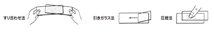
The method of smearing differs depending on the type and properties of the test material. Please smear according to the instructions below.

Specimen fixation

Fixation is the process of stopping changes such as degeneration and lysis of cells, so it is necessary to fix the cells immediately after smear. Wet fixation is for Papanicolaou staining, PAS staining, etc. Dry fixation is for Giemsa staining, etc.
The quality of fixation, along with the collection site and method, is one of the important factors that influences the cytodiagnosis decision. Please fix it quickly.

  1. Wet fixation【Gynecological cytodiagnosis, general cytodiagnosis】

    Fix the smeared glass slide by immersing it in 95% ethanol for at least 30 minutes. 
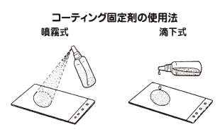
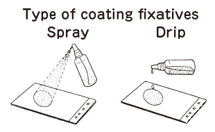
For wet fixation, there is also a coating fixation method using Cytotac, etc.
    If a coating fixative is used, the fixative should be sprayed or dripped thoroughly over the entire surface of the smear to avoid unevenness.

  2. Dry fixation【General cytodiagnosis】

    Immediately after smearing, dry the smeared surface rapidly with a fan or cold air dryer. Natural drying is inadequate as it causes uneven drying.
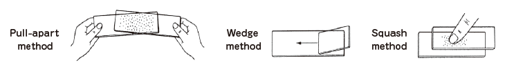
■ Test material, specimen volume

【General cytodiagnosis [Item code: 00541 00541 8]】
  1. Sputum
    Place a "red bean" sized sputum between two glass slides and spread it out evenly, crushing it back and forth, left and right (pull-apart method).
    Since cancer cells are mostly found in the blood sputum, opaque white, and jelly-like mucus areas, smear these areas.
    Specimen volume: Wet-fixed smear specimen 2 slides
  2. Rubbing material (bronchus, digestive tract, pleura/peritoneum, etc.)
    Use a brush or cotton swab to rub the lesion and smear it on a glass slide. The smear should be fixed quickly, as the rubbed material tends to dry out very easily.
    Specimen volume: Wet-fixed smear specimen 1 slide, Dry-fixed smear specimen 1 slide
  3. Liquid material (Urine, pleural fluid, ascites, pericardial fluid, scrotal edema fluid, various washing fluid, etc.)
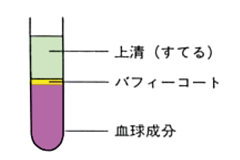
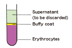
    After centrifugation at 1500 rpm for 5 minutes, smear the sediment by the pull-apart method or the wedge method. For hematological specimens, smear the buffy-coated portion of the specimen, which contains a large amount of nucleic components.
    Specimen volume: Urine, pleural fluid, ascites, pericardial fluid, scrotal edema fluid, various washing solutions, etc. Wet-fixed smear sample 1 slide, Dry-fixed smear sample 1 slide
  4. Needle-puncture aspiration material (Breast, thyroid, lung, lymph node, prostate, testicle, soft part tumor, liver, etc.)
    Please blow the aspirated material into the center of the glass slide and smear gently. If the amount of specimen is very small, peel off the other glass slide after aligning it with the other glass slide to make two slides.
    If a larger amount is obtained, or if the volume is larger, smear the materials using the wedge method.
    Specimen volume: Wet-fixed smear specimen 1 slide, Dry-fixed smear specimen 1 slide
  5. Imprinted specimen (various fixed tumors, lymph nodes, etc.)
    Please lightly imprint the new tissue split surface, cut with a scalpel or razor, on the glass slide.
    Specimen volume: Wet-fixed smear specimen 1 slide, Dry-fixed smear specimen 1 slide
  6. Squash specimen (Central nervous system tumors, thyroid tumors, etc.)
    Please place a small piece of tissue (1-2 mm in diameter) between two glass slides, press it lightly, and when the tissue stretches, remove the glass slide, fix the thicker one with wet-fix method, and fix the thinner one with dry-fix method.
    Specimen volume: Wet-fixed smear specimen 1 slide, Dry-fixed smear specimen 1 slide
<Precaution>

Allowable time from specimen collection to smear fixation (room temperature)

  • Sputum, pleural fluid, ascites, pericardial fluid...within 12 hours
  • Urine, puncture fluid, washing fluid, spinal fluid ... within 1 hour
  • Bile, pancreatic fluid, and duodenal fluid... collected in ice-cold containers, within 1 hour
  • Rubbing material・Inprinted material・squashed material…within 5 second
【Cytodiagnosis (gynecology) [Item code: 00695 00695 8], Cytodiagnosis (Gynecology Bethesda System) [Item code: 06242 66242 5]】
  1. Gynecology material

    Wet fixation should be performed immediately after smear as it tends to dry out easily.
    Specimen volume: Wet-fixed smear specimen 1 slide

  • Please note that the following specimens cannot be requested due to specimen handling.
    • Formalin-fixed specimen
    • Frozen specimen
    • Unrepairable damaged specimens
    • Following test materials
      Tissue, nails, hair, stool, blood, gallstones, stones, bone marrow

Test result reporting

1. Bethesda System

▼Regarding evaluation of suitability of specimens

Suitable specimen: The number of well-preserved, clearly visible squamous epithelium cells should be 8000-12000 for direct smears and at least 5000 for LBCs.
Unsuitable specimen: This applies if 75% or more of the squamous epithelium cells are obscured, if they are covered by inflammatory cells, hemorrhage, or if artifacts due to excessive dryness are seen.

* The following cell determination will be performed if the specimen is deemed suitable.

Bethesda notification Explanation Estimated lesion
Negative (NILM) Negative Microorganisms
Other non-neoplastic findings
Squamous epithelium abnormalities
ASC-US Atypical squamous cells of unknown significance Suspected low-grade squamous intraepithelial lesion
ASC-H Atypical squamous cells that cannot exclude HSIL Suspected high-grade squamous intraepithelial lesion
LSIL Low-grade squamous intraepithelial lesion HPV infection
Low-grade dysplasia
HSIL High-grade squamous intraepithelial lesion Moderate dysplasia
High-grade dysplasia
Intraepithelial carcinoma
SCC Squamous cell carcinoma Squamous cell carcinoma
Glandular abnormalities and other malignancies
AGC Atypical glandular cells Suspected glandular atypia or adenocarcinoma
AIS Adenocarcinoma in situ Adenocarcinoma in situ
Adenocarcinoma Adenocarcinoma Adenocarcinoma
Other malig. Other malignant tumor Other malignant tumor

2. Class determination

Class I
: Absence of atypical or abnormal cells.
Class II
: Atypical cytology but no evidence of malignancy.
Class III
: Cytology suggestive of,but not conclusive for malignancy.
Class IIIa
: Probably benign atypia.
Class IIIb
: Malignancy suspected.
Class IV
: Cytology strongly suggestive of malignancy.
Class V
: Cytology conclusive for malignancy.

* For Class determination, I and II can be read as negative,
III, IIIa, and IIIb as suspicious positive, and IV and V as positive.

3. ABC determination

Classification Cytological findings Instructing category
A No histiocytes in sputum Materials are unsuitable, retest
B Only normal epithelial cell
Basal cell proliferation
Low-grade atypical squamous cells
Ciliated columnar epithelium cells
No abnormality now
Next periodic test
C Moderate atypical squamous cells
Columnar epithelial cells with enlarged nuclei and deep staining
Re-smearing and retest within 6 months
D High-grade (borderline) atypical squamous cells or cells suspicious for malignancy are present Immediately conduct a detailed test
E Malignant tumor cells are present.
Criteria and instruction categories for sputum cytodiagnosis in lung cancer screening (revised 2016) Clinical and Pathological Lung Cancer Treatment Regulations, 8th ed.
Copyright © SRL,Inc. All rights reserved.
  • 株式会社エスアールエル
  • H.U.グループホールディングス
  • SRL, Inc.
  • H.U. Group Holdings, Inc.